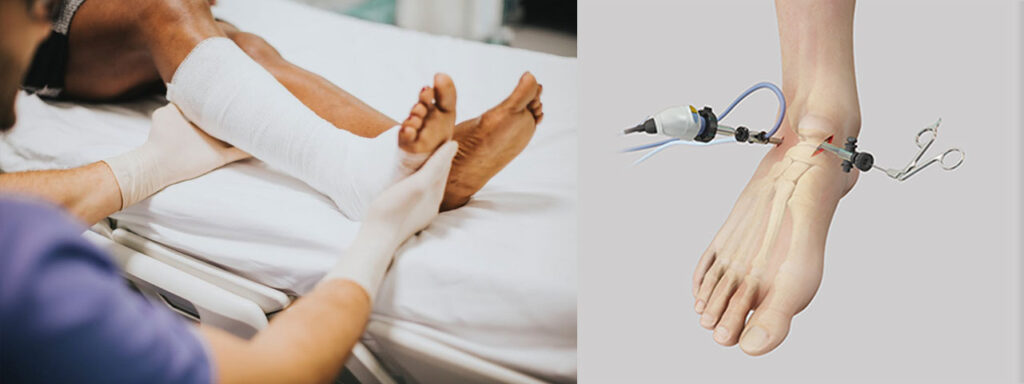
Foot and Ankle Arthroscopy is a minimally invasive surgical procedure used to diagnose and treat various conditions affecting the foot and ankle joints. During this procedure, a small camera called an arthroscope is inserted into the joint through tiny incisions. The camera allows the surgeon to visualize the inside of the joint on a screen, enabling them to identify problems and perform necessary treatments using specialized instruments, all through small openings instead of large, open incisions.
Indications for Foot and Ankle Arthroscopy:
Foot and ankle arthroscopy is typically used for the following conditions:
Ankle Arthritis:
- Osteoarthritis, rheumatoid arthritis, or post-traumatic arthritis (resulting from previous ankle injuries) can lead to pain, stiffness, and swelling in the ankle joint.
- Arthroscopic Treatment: The surgeon can remove damaged tissue, bone spurs, or loose cartilage to improve joint function and reduce pain. In severe cases, arthroscopy may also be used to prepare the joint for ankle replacement surgery.
Ankle Impingement:
- Anterior ankle impingement occurs when bone spurs or soft tissue growth causes the front of the ankle joint to become pinched, often resulting from previous injury.
- Arthroscopic Treatment: The surgeon can remove the bone spurs or excess tissue that cause the impingement, allowing for better joint movement and reduced pain.
Tendon or Ligament Injuries:
- Tendon tears or ligament damage in the ankle can result from sprains or other injuries, causing pain and instability.
- Arthroscopic Treatment: The surgeon may repair torn tendons, remove damaged tissue, or stabilize ligaments to restore function and improve stability.
Loose Bodies in the Ankle:
- Loose bodies (such as fragments of bone or cartilage) can float in the joint and cause pain, swelling, and difficulty moving the ankle.
- Arthroscopic Treatment: These loose bodies can be removed with minimal disruption to the surrounding tissue, improving joint mobility.
Synovitis (Inflammation of Joint Lining):
- Inflammation of the synovial membrane, which lines the joint, can result in pain and swelling.
- Arthroscopic Treatment: The surgeon can clean out the inflamed tissue and remove any scar tissue or debris that may be causing irritation.
Cartilage Damage:
- Damage to the cartilage in the ankle joint can lead to pain, swelling, and decreased mobility.
- Arthroscopic Treatment: Procedures like microfracture or cartilage debridement can be performed to stimulate the growth of new cartilage or remove damaged cartilage, promoting healing.
Tarsal Tunnel Syndrome:
- This condition occurs when the posterior tibial nerve is compressed in the tarsal tunnel (a passageway near the ankle).
- Arthroscopic Treatment: The surgeon can release any structures causing nerve compression, providing relief from pain and numbness.
Haglund’s Deformity:
- A bone enlargement at the back of the heel can cause irritation of the Achilles tendon, leading to pain and inflammation.
- Arthroscopic Treatment: The surgeon may remove the bony prominence and any associated damaged tissue to relieve pressure on the Achilles tendon.
Chronic Ankle Instability:
- Ankle instability occurs when the ligaments are stretched or torn, often from repeated ankle sprains, leading to weakness or frequent ankle rolling.
- Arthroscopic Treatment: Arthroscopy can be used to inspect the ligaments, clean up damaged tissue, and stabilize the joint.
How Foot and Ankle Arthroscopy is Performed:
Preoperative Preparation:
- Consultation: Before surgery, the surgeon will assess your medical history and perform a physical examination. Imaging studies such as X-rays or MRI scans are often used to evaluate the condition of the joint.
- Anesthesia: Foot and ankle arthroscopy is usually performed under general anesthesia or regional anesthesia (such as a nerve block) to numb the area.
- Positioning: The patient’s leg is positioned to give the surgeon clear access to the ankle. The foot and ankle may be placed on a support, with the knee bent or straight, depending on the condition being treated.
Surgical Procedure:
- Incisions: The surgeon makes small incisions, usually one near the front of the ankle and one or two on the side. These incisions are about 1–2 cm in length.
- Inspection: The arthroscope is inserted through one of the incisions, providing a real-time view of the inside of the joint on a monitor. This allows the surgeon to assess the joint’s condition and locate any problems.
- Treatment: Based on the diagnosis, the surgeon may:
- Remove damaged cartilage or bone spurs.
- Repair or remove damaged ligaments or tendons.
- Remove loose bodies or debris.
- Perform procedures like microfracture to stimulate cartilage healing or synovectomy to remove inflamed tissue.
- Closure: After the procedure, the incisions are closed with sutures, and a sterile dressing is applied to the area.
Postoperative Care:
- Recovery Room: After surgery, you will be monitored in a recovery room as the anesthesia wears off. You may experience some discomfort, which can typically be managed with medication.
- Pain Management: Pain relief is usually provided with oral medications, and ice therapy can be applied to reduce swelling and inflammation.
- Bracing or Immobilization: In most cases, the foot and ankle may be immobilized with a splint or a boot to reduce movement and allow healing.
- Elevate: The foot should be kept elevated for several days to help reduce swelling.
Advantages of Foot and Ankle Arthroscopy:
- Minimally Invasive: Because of the small incisions, there is less disruption to the surrounding tissues, resulting in reduced scarring, less pain, and quicker recovery compared to open surgery.
- Faster Recovery: Most patients can return to normal activities more quickly after arthroscopy than after traditional surgery, with less downtime.
- Reduced Risk of Infection: Smaller incisions reduce the risk of infection, a common concern with open surgeries.
- Accurate Diagnosis and Treatment: The arthroscope provides a detailed, real-time view of the joint, helping the surgeon to make a more accurate diagnosis and perform precise treatments.
- Outpatient Procedure: Many foot and ankle arthroscopy procedures can be performed on an outpatient basis, meaning you can often go home the same day.
Risks and Complications:
While foot and ankle arthroscopy is a safe procedure, there are some potential risks, including:
- Infection: Although rare, infections can develop at the incision sites.
- Nerve Injury: The procedure may involve some risk of damaging nearby nerves, potentially leading to temporary or permanent numbness or weakness in the foot or ankle.
- Bleeding: Some bleeding can occur, although this is usually minimal.
- Stiffness: Some patients may experience stiffness in the ankle joint, especially if the joint is not properly rehabilitated post-surgery.
- Blood Clots: Although rare, there is a small risk of developing blood clots, especially if you are immobile for a long period.
- Recurrent Symptoms: In some cases, symptoms may return if the underlying condition is not fully addressed or if the joint does not heal properly.
Recovery and Rehabilitation:
Immediate Postoperative Care:
- After surgery, you will likely need to rest and elevate the foot and ankle to reduce swelling.
- A splint, boot, or brace may be applied to stabilize the joint, and you will likely need crutches for the first few days or weeks to avoid putting weight on the ankle.
Physical Therapy:
- Phase 1 (Initial Recovery): Focus on reducing swelling and pain. Range-of-motion exercises will begin soon after surgery to prevent stiffness.
- Phase 2 (Strengthening): As healing progresses, strengthening exercises will be introduced to rebuild muscle strength and improve stability in the ankle.
- Phase 3 (Return to Activity): Gradually, patients can return to normal activities. For athletes or those with high-demand physical jobs, more advanced exercises may be introduced to restore full function.
Full Recovery: The recovery timeline varies depending on the specific condition treated, but most patients can expect to return to normal activities within 4-6 weeks. If the surgery involved more extensive procedures or if the injury was severe, the recovery period may extend to several months.