Hip Arthroscopy is a minimally invasive surgical procedure used to diagnose and treat a variety of hip joint problems. During hip arthroscopy, a small camera called an arthroscope is inserted through small incisions around the hip joint to view the inside of the joint and perform necessary treatments. This technique allows surgeons to perform surgery with fewer incisions, resulting in less trauma to the surrounding tissues, quicker recovery times, and less pain compared to traditional open surgery.
Indications for Hip Arthroscopy:
Hip arthroscopy can be used to treat a variety of hip problems, including:
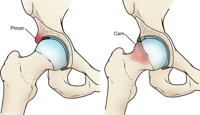
Femoroacetabular Impingement (FAI):
- FAI is a condition where there is abnormal contact between the femoral head (ball) and the acetabulum (socket), leading to joint damage and pain. There are two types of FAI:
- Cam Impingement: A deformity of the femoral head.
- Pincer Impingement: A condition where the acetabulum covers too much of the femoral head.
- Arthroscopic Treatment: Surgeons can trim or reshape the bone to reduce impingement and improve joint movement.
- FAI is a condition where there is abnormal contact between the femoral head (ball) and the acetabulum (socket), leading to joint damage and pain. There are two types of FAI:
Labral Tears:
- The labrum is a ring of cartilage that helps stabilize the hip joint. Tears in the labrum are common and can be caused by trauma or repetitive motion.
- Arthroscopic Treatment: The torn labrum can be repaired or, in some cases, removed if it’s too damaged. In certain cases, the labrum may be reattached using sutures or anchors.
Hip Arthritis:
- Osteoarthritis or other forms of arthritis can cause damage to the cartilage in the hip joint, leading to pain and stiffness.
- Arthroscopic Treatment: In some cases, arthroscopy can be used to remove damaged cartilage or bone spurs, although it is not a cure for arthritis. For more severe cases, hip replacement may be necessary.
Hip Instability:
- Hip instability refers to the inability of the hip joint to stay securely in its socket, causing discomfort and a sensation of the hip “giving way.”
- Arthroscopic Treatment: Surgeons can address soft tissue damage, tighten the joint capsule, or repair ligaments to restore stability.
Snapping Hip Syndrome:
- This condition occurs when tendons or muscles around the hip “snap” over bony prominences, creating a popping or snapping sensation.
- Arthroscopic Treatment: The surgeon can release or reposition the tendons to alleviate the snapping and reduce discomfort.
Cartilage Damage:
- Cartilage in the hip joint can be damaged due to injury, wear and tear, or arthritis.
- Arthroscopic Treatment: Surgeons can remove damaged cartilage, repair the cartilage, or perform procedures like microfracture to stimulate the growth of new cartilage.
How Hip Arthroscopy is Performed:
Preoperative Preparation:
- Consultation: Your surgeon will review your medical history and perform a physical exam. Imaging studies such as X-rays or MRI scans may be ordered to assess the extent of the hip problem.
- Anesthesia: Hip arthroscopy is usually performed under general anesthesia, though regional anesthesia (such as a spinal or epidural block) may be used to numb the lower body.
- Positioning: You will be positioned on the operating table in a way that allows easy access to the hip joint. The leg is often placed in a special traction device to create space in the joint.
Surgical Procedure:
- Incisions: The surgeon will make small incisions (usually about 2-4 incisions, each about 1-2 cm) around the hip joint. One of the incisions is used to insert the arthroscope, while other incisions allow the insertion of specialized surgical instruments.
- Inspection: The surgeon examines the inside of the hip joint using the arthroscope, looking for signs of damage to cartilage, the labrum, or other structures.
- Treatment: Based on the findings, the surgeon can perform a variety of treatments, such as:
- Labral Repair: Reattaching or repairing a torn labrum.
- Bone Reshaping: Removing or reshaping bone spurs or excess bone growth to reduce impingement.
- Cartilage Repair: Repairing or removing damaged cartilage and using procedures like microfracture to promote healing.
- Soft Tissue Repairs: Addressing damaged tendons or ligaments to restore stability and reduce pain.
- Closure: After the procedure is complete, the incisions are closed with sutures, and a sterile dressing is applied to the area.
Postoperative Care:
- Recovery Room: After surgery, you will be taken to a recovery room where you will be monitored as the anesthesia wears off.
- Pain Management: Pain is typically managed with medications, such as NSAIDs, and ice therapy may be used to reduce swelling. Your surgeon may recommend crutches or a hip brace for support initially.
- Physical Therapy: Rehabilitation is crucial for full recovery. The physical therapy program will focus on:
- Restoring range of motion in the hip joint.
- Strengthening the muscles around the hip for stability.
- Gradually returning to normal activities.
Advantages of Hip Arthroscopy:
- Minimally Invasive: Hip arthroscopy requires only small incisions, reducing trauma to the surrounding tissues and minimizing scarring.
- Faster Recovery: Because of the minimally invasive nature, recovery time is typically shorter compared to traditional open surgeries.
- Less Pain: The procedure is associated with less pain and discomfort postoperatively, and pain can often be controlled with medications.
- Fewer Complications: The smaller incisions result in a reduced risk of infection and other complications compared to open surgeries.
- Better Visualization: The arthroscope provides high-definition views of the inside of the joint, allowing the surgeon to make more accurate diagnoses and treatments.
- Outpatient Procedure: Many patients can go home the same day after the surgery, reducing the need for a hospital stay.
Risks and Complications:
Although hip arthroscopy is considered safe, there are potential risks and complications, including:
- Infection: There is a small risk of infection at the incision sites.
- Nerve or Blood Vessel Injury: In rare cases, nerves or blood vessels may be injured during the procedure.
- Stiffness: Some patients may experience stiffness or limited range of motion in the hip joint after surgery.
- Blood Clots: As with any surgery, there is a small risk of developing blood clots in the leg (deep vein thrombosis).
- Hip Dislocation: In rare cases, the hip joint may dislocate during or after surgery, although this is uncommon.
- Continued Pain: Although rare, some patients may continue to experience pain or symptoms after the surgery, especially if the underlying issue wasn’t fully addressed.
Recovery and Rehabilitation:
Immediate Post-Operative Care:
- You will need to rest and elevate your hip in the initial days following surgery to reduce swelling and pain.
- Use of crutches or a walker is common for the first few days or weeks.
- Ice and pain medications will help manage pain and inflammation.
Physical Therapy:
- Phase 1 (First Few Weeks): Focus on reducing pain and swelling, and restoring passive range of motion in the hip joint.
- Phase 2 (3-6 Weeks): Active range-of-motion exercises are introduced, with a focus on strengthening the muscles around the hip joint.
- Phase 3 (6-12 Weeks): More advanced strengthening exercises to restore function and prepare for a gradual return to normal activities.
- Phase 4 (Beyond 3 Months): Return to higher-impact activities such as sports or running, depending on the procedure performed and progress made during rehab.
Full Recovery: Full recovery typically takes around 3 to 6 months, although this can vary depending on the severity of the injury, the type of surgery performed, and the patient’s adherence to rehabilitation.